
When placing a stethoscope on the chest, we don’t listen to the heart “in general.” We target specific areas, each connected to a valve. Placing the bell or diaphragm in the wrong spot is like trying to find a radio station without knowing the frequency. The four cardiac auscultation areas are these frequencies: points where the sound produced by each valve reaches the surface of the chest most clearly.
Why the auscultation area does not correspond to the actual position of the valve
This is the point that confuses most at first. The aortic valve is located in the center of the chest, slightly to the left. Its auscultation area, however, is found at the second right intercostal space, against the edge of the sternum. The discrepancy is explained by the direction of blood flow.
When the left ventricle ejects blood into the aorta, the jet rises to the right before forming the aortic arch. The sound propagates in the direction of flow, not in a straight line from the valve. The stethoscope therefore captures the noise better where the blood current projects the vibrations against the chest wall.
The same principle applies to the other three valves. The mitral valve projects to the apex of the heart (fifth left intercostal space, along the midclavicular line), because the filling flow of the left ventricle directs towards the apex. To understand the 4 cardiac areas, one must think in terms of blood flow, not the raw anatomical location of the valves.
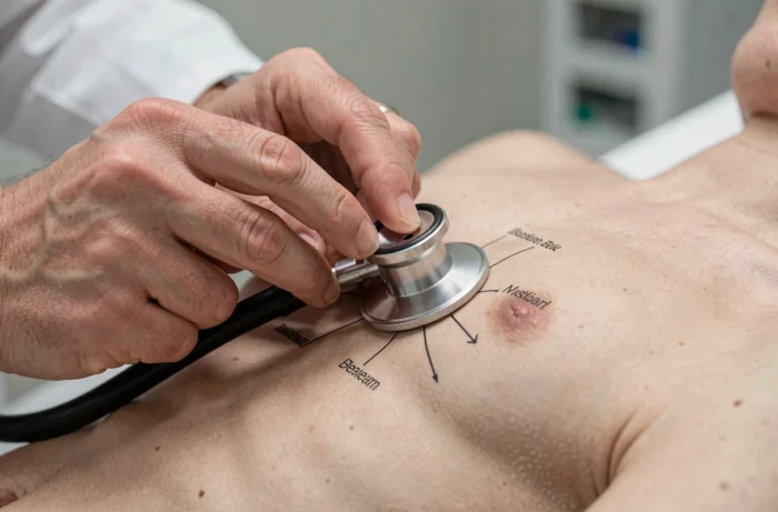
Anatomical landmarks of the four areas on the chest
Before placing the stethoscope, one must know how to count the intercostal spaces. The starting point is the sternal angle (the junction between the manubrium and the body of the sternum), which corresponds to the second intercostal space. From there, you go down rib by rib.
Aortic area
Second intercostal space, right edge of the sternum. This is where the sounds related to the aortic valve are best perceived, including a possible murmur of aortic insufficiency or stenosis. The diaphragm of the stethoscope is suitable for this area, as aortic sounds are rather high-pitched.
Pulmonary area
Second intercostal space, left edge of the sternum. Symmetrical to the aortic area, it captures the sounds of the pulmonary valve. A splitting of the second sound (B2) is often heard better here, especially during deep inspiration.
Tricuspid area
Fourth or fifth intercostal space, left edge of the sternum. This area is dedicated to the tricuspid valve, located between the right atrium and the right ventricle. Tricuspid murmurs intensify during inspiration, as venous return increases the flow in the right chambers. This is a useful clinical clue to distinguish a tricuspid murmur from a mitral murmur.
Mitral area (apex)
Fifth left intercostal space, midclavicular line. It is also called the “apical area.” The patient in the left lateral decubitus position brings the apex closer to the chest wall, which amplifies mitral sounds. The bell of the stethoscope, with light pressure, is particularly suited to capture a low-frequency diastolic rumble.
Diaphragm or bell of the stethoscope: adapting the tool to the area
You may have noticed that certain heart sounds seem to disappear depending on which side of the stethoscope is used? This is not an issue of hearing. The diaphragm filters low frequencies and highlights high-pitched sounds. The bell, on the other hand, captures low sounds as long as you don’t press too hard (excessive pressure stretches the skin and turns it into a diaphragm).
In practice, this distinction changes what you hear at each area:
- Aortic and pulmonary areas: the diaphragm is sufficient in most cases, as ejection murmurs and valve clicks are rather high-pitched sounds.
- Mitral area (apex): start with the bell to search for a B3, B4, or a diastolic rumble, then switch to the diaphragm to listen for a more high-pitched mitral regurgitation murmur.
- Tricuspid area: alternate between the two sides, asking the patient to take a deep breath to accentuate the murmurs from the right heart.

Sequence of cardiac auscultation: building a methodical pathway
Auscultating the four areas in a fixed order prevents forgetting one. The classic sequence starts from the aortic area, descends to the pulmonary area, then the tricuspid area, and ends at the apex (mitral area). Other practitioners prefer to start at the apex. The order matters less than the consistency: always adopting the same pathway anchors the landmarks.
Even before placing the stethoscope, palpating the carotid pulse helps to locate the first heart sound (B1). B1 corresponds to the closure of the mitral and tricuspid valves, at the very beginning of systole. It is almost synchronous with the carotid pulse wave. Identifying B1 using the carotid pulse avoids confusing systole and diastole.
At each area, we first listen for B1 and B2, then look for any added sounds (B3, B4, clicks, rubs) and finally the murmurs. Each step takes a few cardiac cycles. There is no need to rush.
Auscultation simulators and learning cardiac areas
In recent years, next-generation mannequins have integrated the four areas with sounds recorded from real patients, not synthetic noises. These simulators (like the Nasco Auscultation Trainer 2.0 or the SmartScope) offer several dozen thoracic auscultation sites, controlled by tablet.
The main advantage is controlled repetition. The instructor can impose a pathway (aortic, pulmonary, tricuspid, mitral) and vary the pathologies on demand: aortic stenosis murmur, mitral insufficiency, splitting of B2. The recognition of murmurs sharpens through repeated listening, not just theory alone.
Cardiac auscultation remains a clinical gesture where the ear is trained over time. Knowing the projection logic of the valves on the chest, choosing the right side of the stethoscope, and following a systematic pathway are the three pillars of a reliable examination. The rest is a matter of practice, area by area.